Join World's Fastest Growing B2B Network
Join World's Fastest Growing B2B Network


| CAS No. | n/a | Place of Origin | USA |
| Purity | >95% | Classification | Specific Reagents |
| Brand Name | Clonegene | Model Number | CG0110 |
| Application | ELISA, WB, IHC, FFPE, IHC | Packaging | 100 ug/unit |
| Dilution Buffer | 1%BSA/PBS | Form | LIquid |
| Isotype | mouse IgG1 | Activity | Neutralization of PD1/PDL1 interaction |
| Conjugation | Non-conjugated | Concentration | 1mg/ml |
| Specificity | human , mouse PD1 | Clone | J110 |
Programmed cell death protein 1, also known as PD-1 and CD279 (cluster of differentiation 279), is a protein that in humans is encoded by the PDCD1 gene. PD-1 is a cell surface receptor that belongs to the immunoglobulin superfamily and is expressed on T cells and pro-B cells. PD-1 binds two ligands, PD-L1 and PD-L2.
PD-1, functioning as an immune checkpoint, plays an important role in down regulating the immune system by preventing the activation of T-cells, which in turn reduces autoimmunity and promotes self-tolerance. The inhibitory effect of PD-1 is accomplished through a dual mechanism of promoting apoptosis (programmed cell death) in antigen specific T-cells in lymph nodes while simultaneously reducing apoptosis in regulatory T cells (suppressor T cells).
A new class of drugs that block PD-1, the PD-1 inhibitors, activate the immune system to attack tumors and are therefore used to treat cancer.
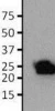
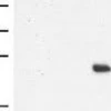
CG0110 is an excellent reagent to investigate the biology of PD-L1
Please note: All products are "FOR RESEARCH USE ONLY AND ARE NOT INTENDED FOR DIAGNOSTIC OR THERAPEUTIC USE"
IgG1
Mouse Monoclonal J110
100 µl of a 1.0 mg/ml preparation
Heat stable, shipped on blue ice. Upon delivery aliquot and store in fridge, longterm storage at -20°C.
ELISA, WB, IHC, FFPE, IHC
One moment please
 Member's Area
 Messages
  Need Help